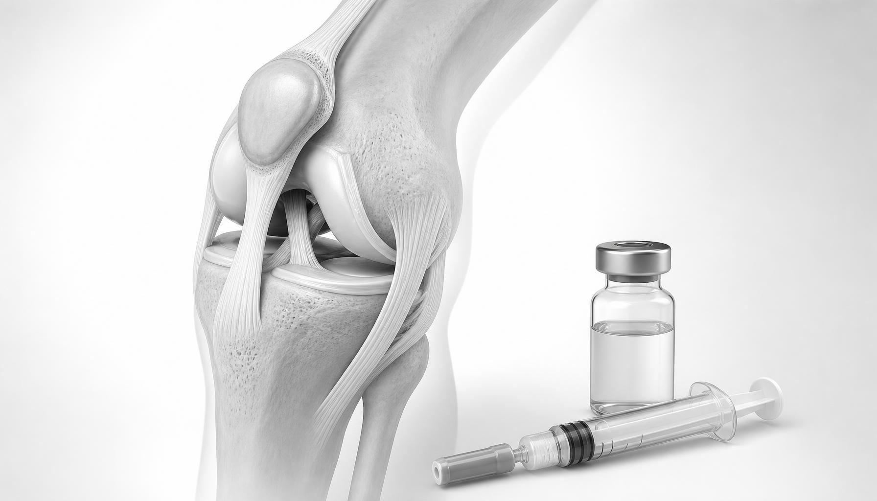
Knee arthritis evaluation in Glastonbury, CT
Dr. Paul Tortland, DO provides comprehensive orthopedic evaluations for patients at his Glastonbury, CT office. The practice sees patients from Glastonbury, Hartford, West Hartford, Farmington, Simsbury, South Windsor, and other Connecticut and New England communities.
📍 59 Sycamore Street, Suite 301, Glastonbury, CT 06033 — Get directions
📞 (860) 430-2821
Learn more about Dr. Tortland · Request an evaluation
Two patients can have nearly identical X-rays of severe knee arthritis and experience completely different levels of pain. The usual explanation, that pain tolerance varies, is only part of the story. A growing body of evidence points to a structural reason: how much of the underlying bone, not just the cartilage, is actually involved.
What X-ray severity does not show about knee arthritis pain
X-rays measure joint space, essentially how much cartilage cushion remains between bones. That's useful, but it's an incomplete picture. X-ray doesn't show what's happening inside the bone itself, where areas of swelling, microdamage, and altered blood flow, called bone marrow lesions, can be present even in joints that look similarly "severe" on X-ray. MRI is required to see them.
Why bone marrow lesions matter in severe knee arthritis
Research has linked bone marrow lesions closely to both pain and disease progression in knee osteoarthritis, often more closely than cartilage loss alone. This helps explain a pattern many patients and physicians notice: someone with "less severe" X-ray findings but a large bone marrow lesion can be in significantly more pain than someone with worse-looking cartilage wear and no lesion. The bone, not just the cartilage, is driving a meaningful share of what the patient feels.
Why standard joint injections miss bone marrow lesions
Standard PRP, BMA, and MFAT injections are placed inside the joint capsule and can meaningfully help cartilage-related and joint-lining pain. But they don't reach tissue inside the bone. For a patient with severe knee arthritis and a confirmed bone marrow lesion, treating the joint alone, however well the injection is prepared, may leave a real source of pain untouched. This is the rationale for intraosseous injection: delivering concentrated PRP or bone marrow aspirate directly into the lesion, typically alongside an intra-articular injection in the same visit.
What studies show about intraosseous injection for knee bone marrow lesions
A systematic review of intraosseous injection for knee bone marrow lesions found the approach safe and beneficial in short-term follow-up, and prospective studies following intraosseous PRP for knee bone marrow lesions have reported sustained improvement in pain and function to a year or more in many patients. The evidence base is still developing, case series and prospective cohorts, not yet large randomized trials, so this is a reasonable option to discuss, not a guaranteed fix.
Who may have a bone marrow lesion driving knee arthritis pain
This is most relevant for patients with moderate-to-advanced knee osteoarthritis whose pain hasn't responded as expected to standard PRP or cellular orthobiologic treatment, and whose MRI confirms a bone marrow lesion. In our practice, we often see this exact pattern: a patient whose pain seems out of proportion to how "mild" their X-ray looks, with an MRI that then reveals a bone marrow lesion driving much of what they're feeling. It's not relevant to every arthritic knee, and it's not a substitute for joint replacement in truly end-stage, bone-on-bone knees with severe deformity.
What bone marrow lesion treatment does not mean
- Not every severe knee has a bone marrow lesion. This explanation applies to some, not all, cases of unexpectedly severe pain.
- Treating a bone marrow lesion does not regrow cartilage.
- It is not a substitute for surgery when a knee is genuinely beyond what injection treatment can help.
- Results vary and aren't guaranteed or necessarily permanent.
Dr. Tortland's Perspective
"The best orthobiologics in the world are worthless without a proper diagnosis," Dr. Tortland has said of his approach to regenerative medicine, and severe knee arthritis is a case in point. When a patient's pain seems out of proportion to their X-ray, or a well-prepared PRP or BMA injection hasn't helped as much as expected, the bone itself is one of the first things he rules in or out — not by guessing, but by looking. Dr. Tortland was in the first group of U.S. physicians registered in musculoskeletal ultrasound by the AIUM, and his "Validate – Investigate – Regenerate" approach starts with confirming exactly what's generating the pain before any injection is planned. MRI, not X-ray, is what answers whether a bone marrow lesion is part of that picture. For a patient with a real lesion, that distinction can be the difference between a treatment plan that's missing a piece and one that finally addresses the whole picture.
Next step: MRI review when knee pain does not match the X-ray
If your knee arthritis pain doesn't seem to match what your X-ray shows, or hasn't responded the way you expected to prior treatment, an MRI review can show whether a bone marrow lesion is part of the picture. Learn more about how intraosseous injection works, or schedule a comprehensive evaluation for an honest answer about your knee.
References on intraosseous treatment of knee bone marrow lesions
- Intraosseous Treatment of Bone Marrow Lesions in the Knee: Surgical Technique. Arthroscopy Techniques, 2024.
- Lychagin A, Lipina M, Garkavi A, et al. Intraosseous injections of platelet rich plasma for knee bone marrow lesions treatment: one year follow-up. International Orthopaedics.
- Centeno C, Cartier C, Stemper I, et al. The treatment of bone marrow lesions associated with advanced knee osteoarthritis: comparing intraosseous and intra-articular injections with bone marrow aspirate and platelet products.